

What Is POCUS in Primary Care?
Point-of-care ultrasound is a focused ultrasound examination performed and interpreted by a trained healthcare professional as part of the patient encounter. Unlike a comprehensive radiology examination, a primary care POCUS assessment is normally designed around a specific clinical question that can be answered within the limits of the operator’s training and the equipment being used.
A focused examination might contribute to a decision such as:
- Whether a patient needs urgent escalation or additional diagnostic imaging
- Whether a superficial finding may contain fluid
- Whether bladder distention or urinary retention should be considered
- Whether ultrasound guidance may improve visualization during vascular access
- Whether a focused musculoskeletal assessment supports the clinical examination
- Whether findings remain indeterminate and require referral or formal imaging
The examination should begin with a clearly defined question. Images should then be acquired using the appropriate preset, probe orientation, depth, gain, and scanning window. Findings must be interpreted in combination with symptoms, history, physical examination, laboratory data, and other available clinical information.
Focused POCUS Applications in Primary Care
The appropriate applications for primary care ultrasound depend on the clinician’s education, supervised training, demonstrated competency, local credentialing requirements, and access to quality assurance. The following examples describe common focused applications and should not be interpreted as permission to perform examinations outside an individual clinician’s scope.
1. Focused Abdominal Assessment
A low-frequency curvilinear or convex probe may be used for selected focused abdominal questions when the operator has appropriate training. Depending on the clinical situation and local protocols, the clinician may assess structures such as the gallbladder, kidneys, urinary bladder, abdominal aorta, or areas where free fluid is clinically relevant.
The purpose is not to perform a complete abdominal ultrasound examination. POCUS may instead identify findings that support a more focused differential diagnosis, help determine urgency, or indicate that comprehensive imaging is necessary.
2. Urinary Bladder and Renal Applications
Focused ultrasound can help trained clinicians evaluate bladder distention and support the assessment of suspected urinary retention. Selected renal views may also be used to look for findings such as collecting-system dilation when appropriate to the clinical question and the operator’s competency.
Ultrasound findings must be interpreted cautiously. Technical limitations, body habitus, bowel gas, incomplete visualization, and operator experience can affect accuracy. Persistent symptoms, abnormal laboratory findings, systemic illness, or concern for obstruction may require formal imaging and specialist evaluation.
3. Lung and Pleural Assessment
Lung ultrasound uses a phased-array, curvilinear, or linear probe to evaluate focused pleural and pulmonary patterns. In properly trained hands, the examination may contribute information about pleural sliding, focal interstitial patterns, consolidation patterns, or pleural fluid.
Lung POCUS should not be interpreted in isolation. Findings depend on the patient’s symptoms, pretest probability, scanning protocol, image quality, and operator experience. Serious cardiopulmonary symptoms require timely escalation and appropriate diagnostic evaluation.
4. Focused Cardiac Views
Limited focused cardiac ultrasound may provide selected information about global cardiac activity, pericardial fluid, and gross ventricular appearance. This application requires dedicated training and should remain clearly distinguished from comprehensive echocardiography.
Primary care clinicians should use focused cardiac imaging only within an established competency and governance framework. Abnormal, uncertain, or clinically discordant findings require formal echocardiography, specialist interpretation, or urgent escalation as appropriate.
5. Superficial Soft-Tissue Assessment
A high-frequency linear probe can display superficial soft-tissue structures and may assist with focused evaluation of a localized area of swelling, redness, pain, or suspected fluid collection. Ultrasound may show whether a finding appears primarily fluid-filled, solid, edematous, or otherwise indeterminate.
Ultrasound appearance alone should not be used to rule out serious infection, malignancy, vascular disease, or another important condition. The patient’s history, examination, systemic symptoms, and need for drainage, laboratory testing, formal imaging, or referral remain essential.
6. Musculoskeletal Applications
A linear probe may be used for focused assessment of superficial tendons, muscles, joints, bursae, and other musculoskeletal structures. POCUS can supplement the physical examination, provide dynamic visualization, and help identify whether additional diagnostic imaging or referral may be appropriate.
Musculoskeletal ultrasound has a significant learning curve. The operator must understand normal anatomy, anisotropy, probe orientation, dynamic maneuvers, and common artifacts. Suspected fracture, infection, neurovascular compromise, severe injury, or unexplained persistent symptoms may require radiography, MRI, specialist assessment, or urgent care.
7. Ultrasound-Guided Vascular Access
A high-frequency linear probe can visualize superficial vessels and surrounding structures during vascular access. Ultrasound guidance may assist with vessel identification, needle visualization, and avoidance of adjacent anatomy when performed by a trained clinician using an appropriate sterile or aseptic technique.
The operator should follow established infection-control practices, use suitable probe covers and sterile gel when indicated, confirm vessel characteristics, and understand the limitations of both short-axis and long-axis needle approaches.
8. Selected Women’s Health and Pelvic Applications
Selected pelvic or women’s health applications may be appropriate for clinicians with specific training, competency, appropriate equipment, consent procedures, infection-control protocols, and institutional authorization. An endocavity examination should never be performed without the required education, patient consent, chaperone and privacy procedures where applicable, and adherence to local policies.
Pain, bleeding, pregnancy-related concerns, hemodynamic instability, or uncertain findings may require urgent laboratory evaluation, formal imaging, emergency assessment, or specialist referral. A limited primary care examination must not delay necessary care.
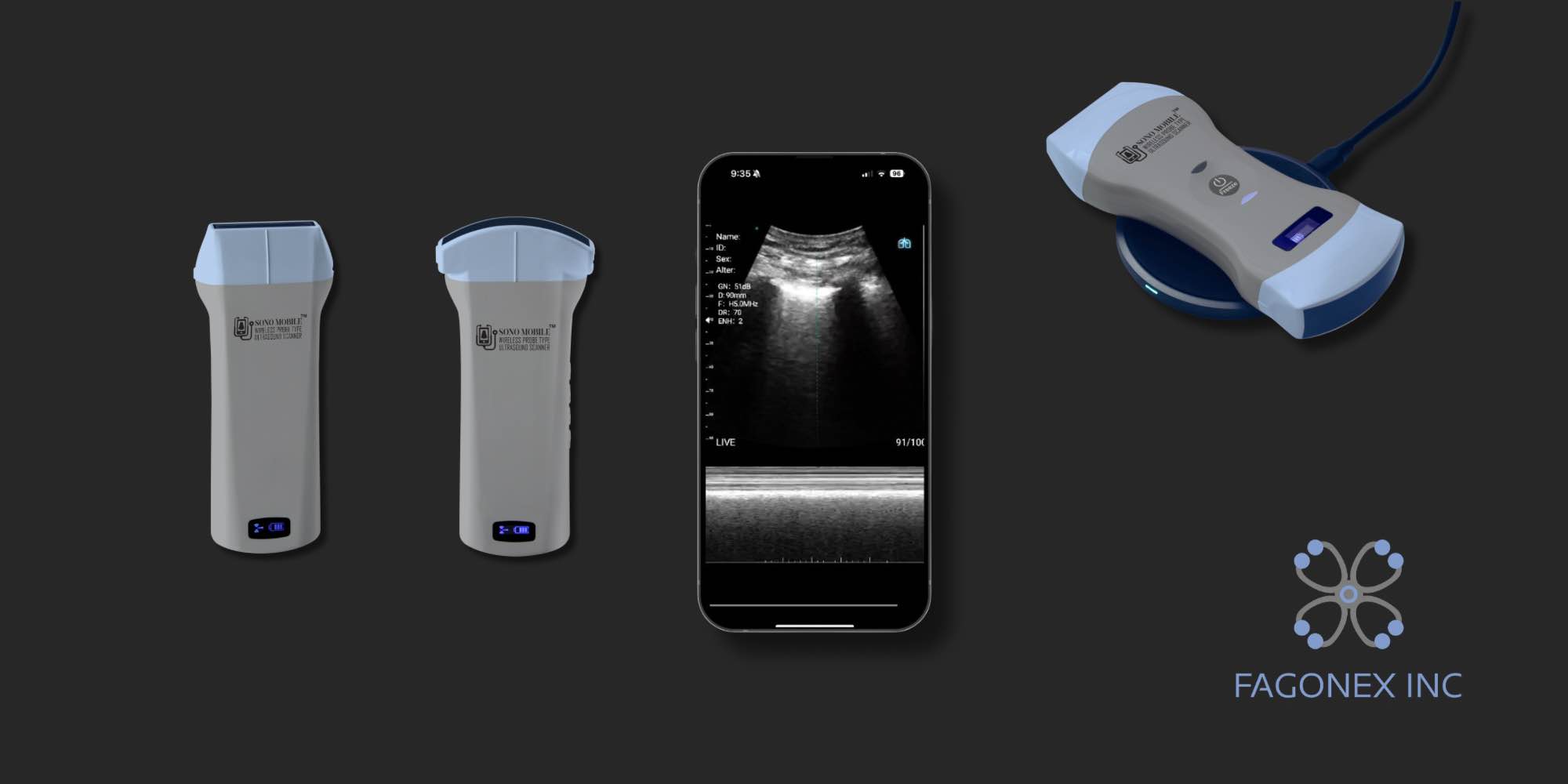
Handheld Ultrasound Probe Selection for Primary Care
Probe selection should be based on the clinical application rather than choosing a device solely because it includes the greatest number of features. The correct probe frequency, footprint, preset, image depth, and ergonomics are important for obtaining usable images.
| Model | Probe configuration | Potential primary care applications | Best suited for |
|---|---|---|---|
| Sono Mobile® CL64 | Dual-head convex and linear configuration with a phased-array preset | Focused abdominal, renal, bladder, lung, vascular-access, superficial soft-tissue, musculoskeletal, and selected focused cardiac workflows | Practices seeking broad application coverage from one handheld device |
| Sono Mobile® C6 | Convex probe | Focused abdominal, renal, urinary bladder, and deeper general imaging applications | Practices primarily performing abdominal and deeper-structure examinations |
| Sono Mobile® L4 | Linear probe | Vascular access, superficial soft tissue, musculoskeletal structures, and other shallow imaging applications | Clinics focusing on superficial anatomy, procedures, and vascular access |
| Sono Mobile® CT61 | Dual-head convex and endocavity configuration | Focused abdominal and selected pelvic or women’s health applications | Appropriately trained clinicians with an established pelvic-imaging workflow and required governance procedures |
Sono Mobile® CL64: Broad Primary Care Versatility
For a primary care practice that expects to scan both superficial and deeper anatomy, the Sono Mobile® CL64 provides the broadest suggested configuration among these models. Its convex and linear scanning options can support abdominal, renal, bladder, lung, vascular-access, soft-tissue, and musculoskeletal workflows, while its phased-array preset may support selected focused cardiac applications.
Versatility does not eliminate the need for application-specific training. Each examination requires different anatomy knowledge, image-acquisition skills, documentation standards, and competency assessment.
Sono Mobile® C6: Abdominal and General Imaging
The Sono Mobile® C6 is a convex model suited to deeper imaging. It may be considered when the practice’s primary use cases include the abdomen, urinary bladder, kidneys, and other general-imaging applications that benefit from a wider field of view and greater penetration.
Sono Mobile® L4: Superficial Structures and Procedures
The Sono Mobile® L4 is a linear model suited to superficial structures. Potential applications include vascular access, soft-tissue assessment, musculoskeletal imaging, and visualization of shallow anatomy.
Sono Mobile® CT61: Selected Pelvic Workflows
The Sono Mobile® CT61 combines convex and endocavity scanning options. It may be relevant to appropriately trained clinicians who perform selected abdominal, pelvic, or women’s health examinations within a formal competency, consent, infection-control, privacy, and quality-assurance framework.
View available models in the Sono Mobile® handheld ultrasound product collection. For a more detailed purchasing framework, including portability, compatibility, workflow, support, and total-cost considerations, read How to Choose the Best POCUS Device for Primary Care.
A Practical Primary Care POCUS Workflow
- Define the clinical question.
Decide exactly what the examination is intended to evaluate. Avoid beginning with a broad request to “scan everything.” - Confirm that the examination is within scope.
Ensure the application is covered by the operator’s training, demonstrated competency, credentials, and local policies. - Select the appropriate probe and preset.
Match probe frequency, footprint, penetration, and preset to the target anatomy and clinical question. - Explain the examination and obtain consent.
Provide an appropriate explanation and follow local consent, privacy, chaperone, and documentation requirements. - Acquire images systematically.
Use a defined scanning protocol and optimize depth, gain, focus, orientation, and patient positioning. - Interpret findings within the clinical context.
Combine ultrasound observations with history, examination, vital signs, laboratory results, and pretest probability. - Classify the result.
Document whether the examination is positive, negative, limited, technically inadequate, or indeterminate for the focused question. - Document and store images when required.
Follow organizational policies for image retention, patient identifiers, privacy, reports, quality review, and billing. - Escalate when necessary.
Arrange formal imaging, specialist consultation, emergency evaluation, or follow-up when the examination is abnormal, incomplete, uncertain, or inconsistent with the clinical presentation.

Training, Quality Assurance, and Ultrasound Safety
Competency Is Application-Specific
Competency in one POCUS application does not automatically establish competency in another. For example, experience with vascular access does not by itself establish competency in focused cardiac, lung, abdominal, pelvic, or musculoskeletal ultrasound.
A structured training pathway may include:
- Foundational ultrasound physics and instrumentation
- Normal and abnormal anatomy
- Probe selection and orientation
- Image optimization and artifact recognition
- Supervised image acquisition
- Interpretation in the appropriate clinical context
- Documentation and image archiving
- Competency assessment for each clinical application
- Ongoing image review and quality assurance
- Defined pathways for indeterminate or discordant results
Quality Assurance
A primary care POCUS program should include regular review of image quality, interpretation, documentation, clinical integration, and follow-up outcomes. Quality review can help identify recurring problems involving probe orientation, depth, gain, incomplete scanning protocols, artifacts, overinterpretation, or insufficient escalation.
Practices should establish clear policies covering:
- Who is authorized to perform each examination
- How competency is assessed and maintained
- Which images must be saved
- How findings are documented
- How patient data are protected
- How probes and accessories are cleaned and disinfected
- How discrepant findings and adverse events are reviewed
- When formal diagnostic imaging is required
Responsible Use of Diagnostic Ultrasound
Medical ultrasound does not use ionizing radiation, but it should still be used responsibly. Operators should follow the principle of using the lowest output and shortest examination time reasonably needed to obtain the required diagnostic information. Unnecessary scanning should be avoided.
For additional safety information, review the U.S. Food and Drug Administration’s ultrasound imaging guidance and the American Institute of Ultrasound in Medicine official statements.
Cleaning, Disinfection, and Infection Control
Probes and accessories should be cleaned and disinfected according to the manufacturer’s instructions, the type of examination, infection-control standards, and local policies. Endocavity probes require particularly careful handling, appropriate probe covers, and the required level of disinfection between patients. A probe cover does not replace required cleaning and disinfection.
Limitations of Primary Care Ultrasound
POCUS is highly dependent on the operator, patient, clinical question, equipment, and scanning environment. Image quality may be limited by body habitus, pain, wounds, dressings, bowel gas, patient position, inability to cooperate, or the depth and location of the target anatomy.
Important limitations include:
- A focused examination evaluates only a limited question
- Incomplete visualization can create false reassurance
- Artifacts may imitate or obscure pathology
- Early or subtle disease may not produce recognizable findings
- A normal focused scan may not exclude serious illness
- Unexpected findings may require formal characterization
- Image interpretation depends on training and experience
When Formal Imaging or Referral May Be Necessary
Formal diagnostic imaging, specialist consultation, or urgent escalation should be considered when:
- The POCUS result is technically limited or indeterminate
- The ultrasound finding does not explain the clinical presentation
- Symptoms persist or worsen despite a reassuring focused examination
- A serious condition remains clinically possible
- A comprehensive study, measurement, Doppler assessment, or specialist interpretation is required
- An unexpected mass, structural abnormality, or complex finding is identified
- The patient has unstable vital signs or other emergency features