
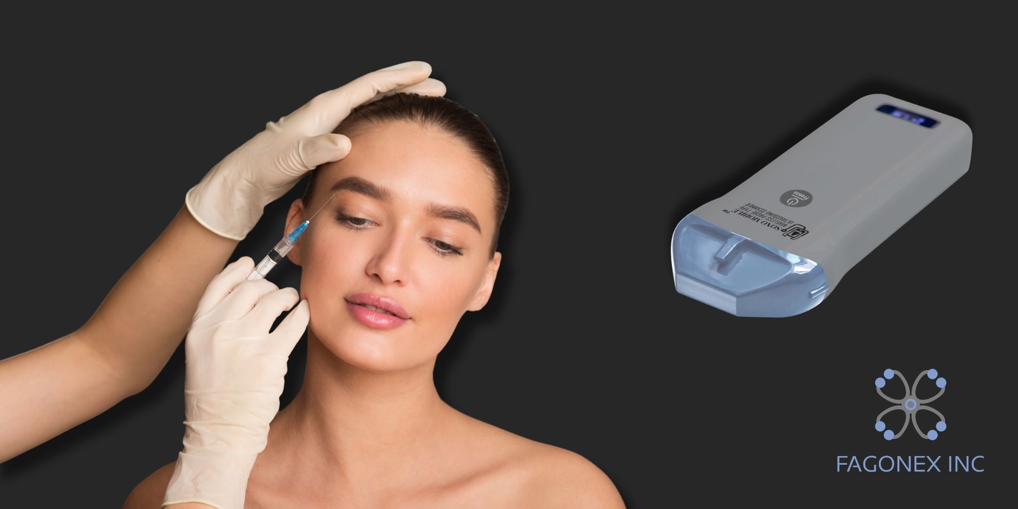
Aesthetic Ultrasound with Sono Mobile® L2: Precision Imaging for Filler Assessment and Treatment Guidance
Enhance Precision and Safety in Aesthetic Procedures with Sono Mobile® L2
Aesthetic ultrasound is becoming an increasingly valuable imaging tool for physicians who need a clearer understanding of facial anatomy, soft-tissue structures, and the location of previously injected products. Sono Mobile® L2 is a compact, high-frequency handheld ultrasound designed to help qualified healthcare professionals visualize relevant anatomical structures and assess soft tissues in real time.
By providing dynamic ultrasound imaging at the point of care, Sono Mobile® L2 can support physicians before, during, and after aesthetic procedures, including dermal filler assessment, hyaluronidase treatment, facial anatomy evaluation, complication assessment, and treatment follow-up.
For physicians performing injectable aesthetic procedures, ultrasound can provide valuable information that may not be available through visual examination or palpation alone.
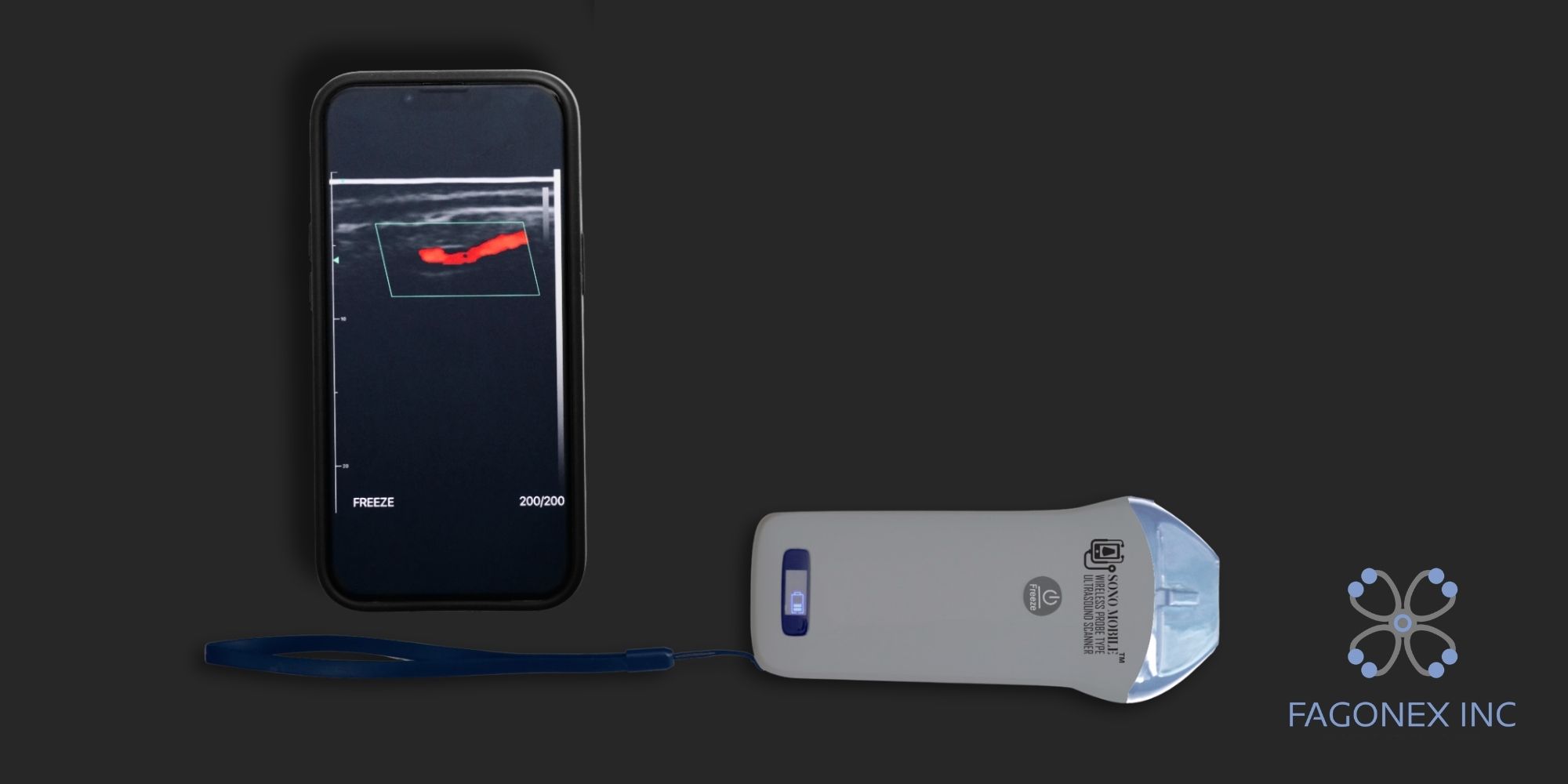
How Aesthetic Ultrasound with Sono Mobile® L2 Supports Clinical Practice
Precision and Safety During Injectable Procedures
Ultrasound imaging allows physicians to visualize facial soft tissues and relevant anatomical structures in real time. During aesthetic procedures, this additional information can support more informed treatment planning by helping identify blood vessels, tissue layers, and previously injected filler deposits.
For procedures involving dermal fillers, including hyaluronic acid fillers, Sono Mobile® L2 can complement clinical assessment and help qualified healthcare professionals make more informed decisions regarding treatment location, depth, and technique.
Dermal Filler Localization and Distribution Assessment
One of the most important applications of aesthetic ultrasound is the localization and assessment of previously injected dermal fillers.
Sono Mobile® L2 can help physicians evaluate the location, depth, and distribution of hyaluronic acid filler within facial soft tissues. This can be particularly useful when assessing:
- Overfilled areas
- Uneven filler distribution
- Filler migration
- Filler placed at an unexpected depth
- Persistent or recurrent filler deposits
- Palpable nodules or irregularities
- Asymmetry following previous treatments
- Complications associated with injectable procedures
Ultrasound findings can help determine whether an observed irregularity corresponds to dermal filler or another soft-tissue finding. This information can support a more individualized and targeted treatment strategy.
Ultrasound-Guided Hyaluronidase Treatment and Follow-Up
When hyaluronic acid filler needs to be dissolved, ultrasound can provide valuable information before, during, and after hyaluronidase treatment.
Sono Mobile® L2 can help physicians identify the location and extent of the filler and evaluate its relationship to surrounding anatomical structures. This may support a more targeted approach, particularly when filler deposits are difficult to identify through visual examination or palpation alone.
Following hyaluronidase administration, ultrasound can also be used to reassess the treated area, document changes, and determine whether residual filler remains. Additional clinical evaluation or treatment may then be considered when appropriate.
Clinical note: Ultrasound should be used as an adjunct to clinical assessment by appropriately trained healthcare professionals. Decisions regarding hyaluronidase administration must follow established clinical protocols, applicable regulations, and professional judgment.
Improved Anatomical Awareness and Reduced Reliance on Blind Injection
Facial anatomy varies significantly between patients, and previous aesthetic treatments can further alter or complicate the anatomical picture.
Facial ultrasound provides real-time visualization beneath the skin, helping physicians better understand the individual patient’s anatomy before and during an aesthetic procedure. It may help identify tissue layers, filler deposits, and relevant vascular structures that cannot be reliably evaluated through surface anatomy or palpation alone.
By providing additional imaging information, Sono Mobile® L2 can support more informed procedural planning and reduce reliance on blind injection techniques.
Real-Time Imaging During Aesthetic Treatment
Unlike a static image, ultrasound provides dynamic, real-time visualization of facial anatomy and soft tissues.
During selected aesthetic procedures, physicians can use Sono Mobile® L2 to observe relevant structures and filler deposits as the examination or treatment progresses. This allows ultrasound findings to be considered alongside the patient’s clinical presentation.
After treatment, ultrasound may also help assess changes in the appearance, location, or distribution of filler and support decisions regarding follow-up care.
Pre-Treatment Planning and Long-Term Follow-Up
Effective aesthetic treatment begins with a clear understanding of the patient’s anatomy and previous treatment history.
Before performing injectable procedures, Sono Mobile® L2 can support the assessment of facial soft tissues and previously injected products. This may be especially valuable for patients who have undergone multiple aesthetic treatments or whose previous filler type, location, or depth is uncertain.
Aesthetic ultrasound may support treatment planning and follow-up involving:
- Facial filler procedures
- Lip augmentation
- Filler correction
- Hyaluronidase treatment planning
- Facial rejuvenation
- Filler complication assessment
- Post-procedure swelling
- Soft-tissue changes
- Selected fat-grafting and post-liposuction assessments
Documenting ultrasound findings over time can also contribute to more consistent patient records and support continuity of care.
Non-Invasive Assessment of Aesthetic Complications
Patients may present with visible or palpable abnormalities following previous aesthetic procedures. Depending on the clinical situation, ultrasound can provide a non-invasive method for assessing selected soft-tissue findings, including:
- Nodules
- Cysts
- Localized swelling
- Filler deposits
- Soft-tissue irregularities
- Suspected filler migration
- Changes following injectable procedures
By combining imaging findings with medical history and physical examination, physicians can develop a more informed understanding of the affected area and determine whether further evaluation, treatment, or referral may be appropriate.
Support in Time-Sensitive Complication Assessment
Certain complications associated with aesthetic procedures require immediate professional assessment and management.
When used by appropriately trained clinicians, a handheld ultrasound can provide real-time information about vascular anatomy and blood flow. In suspected vascular complications, this information may support clinical decision-making and help identify relevant anatomical structures during ultrasound-guided assessment or intervention.
When hyaluronidase is clinically indicated for a complication involving hyaluronic acid filler, ultrasound may provide additional information for treatment planning and subsequent follow-up.
Important: Ultrasound does not replace established emergency protocols, clinical judgment, or immediate medical management. Suspected vascular occlusion and other serious complications require urgent assessment and treatment by qualified healthcare professionals.

Portable Imaging, Patient Communication, and Clinical Education
As a wireless handheld ultrasound system, Sono Mobile® L2 allows physicians to bring real-time imaging directly to the patient without requiring a dedicated imaging room or a conventional cart-based ultrasound system.
This makes Sono Mobile® L2 suitable for qualified healthcare professionals working in:
- Aesthetic medicine clinics
- Dermatology practices
- Plastic surgery clinics
- Cosmetic medicine centers
- Hospitals
- Medical spas operating under qualified medical supervision
- Clinical training and educational environments
For facial ultrasound examinations, appropriate ultrasound gel supports effective acoustic coupling between the probe and the patient’s skin. When barrier protection is required, compatible disposable probe covers can help support probe hygiene and infection-control practices in accordance with applicable clinical protocols.
Real-time imaging can also help physicians explain facial anatomy, filler location, and treatment considerations to patients. Showing relevant ultrasound findings during a consultation may support clearer communication and help patients better understand their anatomy and proposed treatment plan.
Sono Mobile® L2 can additionally serve as an educational tool for physicians learning facial anatomy and ultrasound-guided aesthetic procedures. Live imaging helps trainees understand the relationship between the skin, subcutaneous tissue, blood vessels, filler deposits, and deeper anatomical structures.
In multidisciplinary aesthetic care, ultrasound findings may also support documentation and communication among aesthetic physicians, dermatologists, plastic surgeons, emergency physicians, and other qualified healthcare professionals involved in assessment, referral, treatment, and follow-up.
Why Choose Sono Mobile® L2 for Aesthetic Ultrasound?
Sono Mobile® L2 brings point-of-care ultrasound into the hands of aesthetic healthcare professionals in a compact and convenient format.
Its potential applications extend beyond routine imaging. In aesthetic medicine, ultrasound can become an important component of a physician’s clinical workflow; from pre-treatment assessment and filler localization to complication evaluation, hyaluronidase treatment planning, and post-treatment follow-up.
Key Benefits of Sono Mobile® L2 in Aesthetic Medicine
- Real-time ultrasound imaging
- Assessment of facial soft-tissue anatomy
- Filler localization and distribution assessment
- Support for hyaluronic acid filler evaluation
- Ultrasound-assisted treatment planning
- Assessment of aesthetic complications
- Support for hyaluronidase treatment planning
- Post-treatment follow-up
- Portable point-of-care imaging
- Patient education and communication
- Clinical training and education
- Convenient use in aesthetic clinics and medical practices
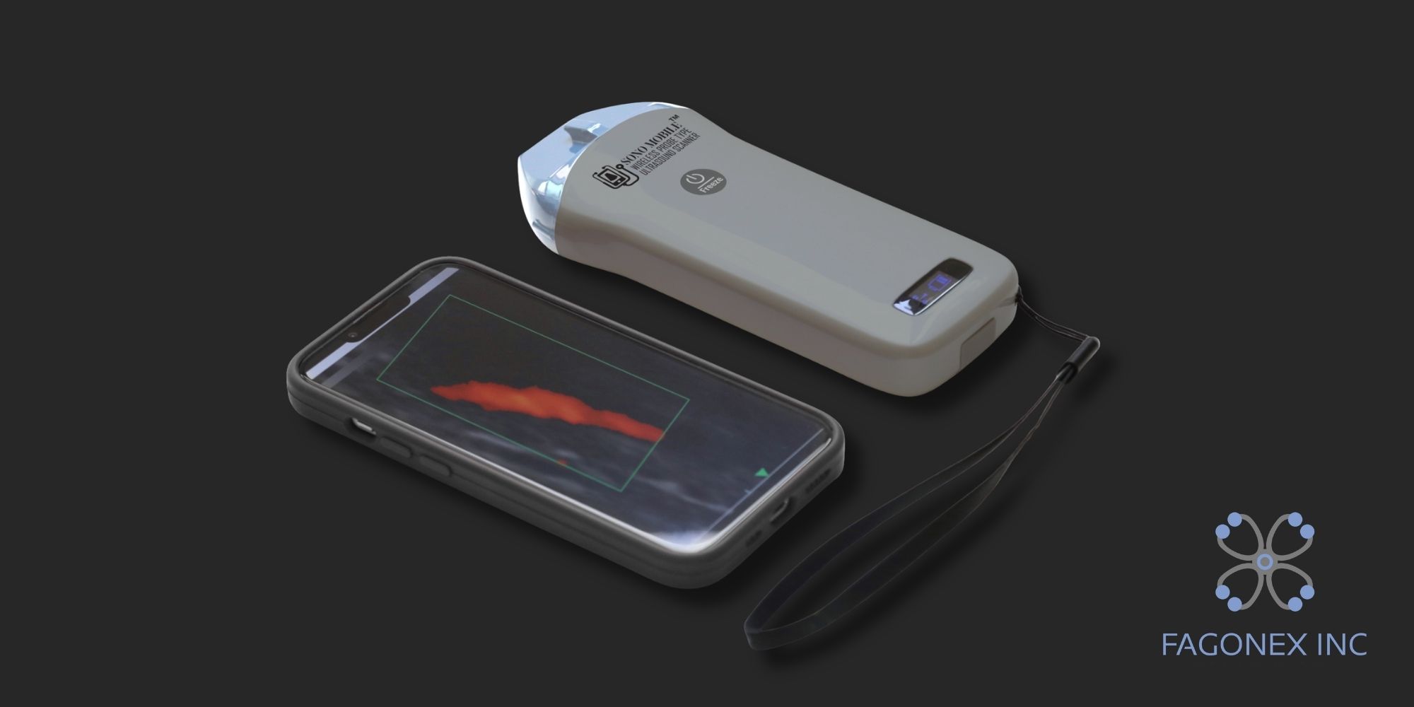
Sono Mobile® L2: Bringing Ultrasound Closer to Aesthetic Care
For aesthetic physicians, having access to ultrasound at the point of care can provide an additional layer of information that complements physical examination and clinical expertise.
From identifying the location of existing filler to evaluating soft-tissue findings and supporting treatment planning, Sono Mobile® L2 can help physicians incorporate real-time imaging into their aesthetic workflow.
Whether you are looking to improve filler assessment, ultrasound-guided aesthetic procedures, hyaluronidase treatment planning, complication evaluation, or patient communication, Sono Mobile® L2 provides a portable solution designed to bring ultrasound imaging directly to the point of care.
Explore Sono Mobile® L2 in Aesthetic Medicine
See how Sono Mobile® L2 can support your aesthetic practice with portable, real-time ultrasound imaging.
Book a personalized demonstration today and discover how Sono Mobile® L2 can help you integrate point-of-care ultrasound into your clinical workflow.
Request Your Sono Mobile® L2 Demo
Contact us today to schedule a personalized demonstration and explore the possibilities of handheld ultrasound for aesthetic medicine.
Clinical applications and use of ultrasound-guided procedures should be performed by appropriately trained and qualified healthcare professionals and in accordance with applicable clinical guidelines, regulations, and device indications.
Editorial Information
Prepared by the FAGONEX Clinical Content Team
Last updated: August 2026
This content is provided for professional educational and informational purposes. It does not constitute medical advice and does not replace clinical training, professional judgment, established emergency protocols, or applicable clinical guidelines. Ultrasound examinations and ultrasound-guided procedures should be performed by appropriately trained and qualified healthcare professionals in accordance with local regulations and the device’s approved indications for use.
Scientific References
- Urdiales-Gálvez F, et al. Ultrasound Patterns of Different Dermal Filler Materials Used in Aesthetics. Journal of Cosmetic Dermatology. 2021.
- Mlosek RK, et al. The Use of High-Frequency Ultrasonography for the Diagnosis of Palpable Nodules After the Administration of Dermal Fillers. Journal of Ultrasonography. 2020.
- Schelke LW, Velthuis P, Kadouch J, Swift A. Early Ultrasound for Diagnosis and Treatment of Vascular Adverse Events with Hyaluronic Acid Fillers. Journal of the American Academy of Dermatology. 2023.
- Shekarriz P, et al. Ultrasound-Assisted Management of Filler-Related Complications. Journal of Cosmetic Dermatology. 2022.
- Boey JJE, et al. Superior Outcomes with Ultrasound-Guided Hyaluronidase for Impending Filler-Induced Facial Skin Necrosis: A Systematic Review and Pilot Meta-Analysis. Aesthetic Plastic Surgery. 2025.
- U.S. Food and Drug Administration. Ultrasound Imaging: Uses, Benefits, Risks, and Information for Healthcare Providers.
See Sono Mobile® L2 in Action
Explore how Sono Mobile® L2 can bring real-time ultrasound imaging to aesthetic medicine. In the videos below, see ultrasound scans of different areas of the face and discover how imaging can help physicians visualize facial anatomy, soft tissues, and previously injected dermal fillers.
Watch the scans and explore the possibilities of point-of-care ultrasound in aesthetic medicine. Learn more about how Sono Mobile® L2 can support facial assessment, filler evaluation, treatment planning, and follow-up in aesthetic practices.
Explore the videos below and see Sono Mobile® L2 in action.